- Advanced Stage Hepatocellular Carcinoma Successfully Treated with Transarterial Radioembolization and Multi-tyrosine Kinase Inhibitor Therapy
-
Myung Ji Goh, Wonseok Kang, Dong Hyun Sinn, Geum-Youn Gwak, Yong-Han Paik, Moon Seok Choi, Joon Hyeok Lee, Kwang Cheol Koh, Seung Woon Paik
-
J Liver Cancer. 2020;20(2):160-166. Published online September 30, 2020
-
DOI: https://doi.org/10.17998/jlc.20.2.160
-
-
3,560
Views
-
133
Downloads
-
 Abstract Abstract
 PDF PDF
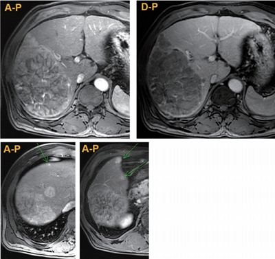
- Transarterial radioembolization (TARE) with yttrium-90 microspheres has become widely utilized in managing hepatocellular carcinoma (HCC). The utility of TARE is expanding with new insights through experiences from real-world practice and clinical trials, and recently published data suggest that TARE in combination with sorafenib may improve the overall survival in selected patients. Here, we report a case of advanced stage HCC that was successfully treated with TARE and sorafenib. The patient achieved complete response (CR) at 12 months after the initial treatment with TARE and sorafenib, followed by additional transarterial chemoembolization and proton beam therapy for local tumor recurrence at 19-month post-TARE. The patient was followed up every 3 months thereafter and still achieved CR both biochemically and radiologically for the following 12 months. A combination strategy of TARE and systemic therapy may be a useful alternative treatment option for selected patients with advanced stage HCC.
- Radiation-induced Myositis after Proton Beam Therapy to Huge Hepatocellular Carcinoma
-
Jihye Kim, Gyu Sang Yoo, Dong Hyun Sinn, Hee Chul Park, Kwang Cheol Koh
-
J Liver Cancer. 2019;19(2):136-142. Published online September 30, 2019
-
DOI: https://doi.org/10.17998/jlc.19.2.136
-
-
6,176
Views
-
119
Downloads
-
2
Citations
-
Abstract
PDF
- Proton beam therapy (PBT) is one of the advances in radiotherapy techniques, which enables dose escalation with lower probability of radiation-induced liver or gastrointestinal injuries. However, the chest wall proximal to the tumor can be affected by high dose irradiation. Here, we report on a 58-year-old male patient who presented with huge hepatocellular carcinoma, received treatment with transarterial chemoembolization and PBT, and developed severe chest wall pain due to radiation-induced myositis. The patient’s symptoms were controlled by oral steroids.
-
Citations
Citations to this article as recorded by  - Pectoralis Major Radiation Myonecrosis After Lung Stereotactic Body Radiation Therapy
Jason Gurewitz, Anand Mahadevan, Benjamin T. Cooper
Practical Radiation Oncology.2023;[Epub] CrossRef - Current role of proton beam therapy in patients with hepatocellular carcinoma
Gyu Sang Yoo, Jeong Il Yu, Hee Chul Park
International Journal of Gastrointestinal Intervention.2021; 10(4): 175. CrossRef
- Cause of Mortality for Hepatocellular Carcinoma Patients who were Diagnosed within the Milan Criteria
-
Hyun-Woo Lee, Dong Hyun Sinn, Wonseok Kang, Geum-Youn Gwak, Yong-Han Paik, Moon Seok Choi, Joon Hyeok Lee, Kwang Cheol Koh, Seung Woon Paik
-
J Liver Cancer. 2016;16(2):101-107. Published online September 30, 2016
-
DOI: https://doi.org/10.17998/jlc.16.2.101
-
-
1,464
Views
-
23
Downloads
-
7
Citations
-
Abstract
PDF
- Background/Aim
s: Hepatocellular carcinoma (HCC) is a unique condition where the cause of
death might not only be due to progressive cancer, but also from liver failure. We evaluated
specific causes of death for HCC patients who were initially diagnosed within the Milan criteria.
Methods
A retrospective cohort of 147 patients with mortality who were initially diagnosed
with HCC within the Milan criteria between January 2008 and December 2012 at a single
institution was reviewed.
Results
During follow-up, 104 patients (70.7%) experienced one or more cirrhotic complications,
such as ascites, variceal bleeding, or hepatic encephalopathy. Near mortality, cancer progression
(exceeding the Milan criteria) was recorded for 102 patients (69.3%), while cirrhosis progression
(greater than two-point increase in Child-Pugh score) was noted in 110 (74.8%) patients. Alphafetoprotein,
protein-induced by vitamin K antagonist-II levels and treatment modality were
associated with cancer progression, while age and Child-Pugh class were associated with
cirrhosis progression. There were 61 patients with in-hospital mortality; cancer progression
plus liver failure was noted in 34 patients (55.7%), liver failure without cancer progression was
seen in 20 patients (32.8%), and only four patients (6.6%) showed mortality from extrahepatic
metastasis without liver failure.
Conclusions
Among HCC patients who were diagnosed within the Milan criteria, most of them
had cirrhosis progression near mortality, and significant proportion died without uncontrolled
cancer growth, mainly due to liver failure. These findings show the importance of liver function
that should be considered in managing HCC patients.
-
Citations
Citations to this article as recorded by - 2022 KLCA-NCC Korea practice guidelines for the management of hepatocellular carcinoma
Journal of Liver Cancer.2023; 23(1): 1. CrossRef - Clinical Outcomes of Hepatitis B Virus–Related Hepatocellular Carcinoma Patients with Undetectable Serum HBV DNA Levels
Jong-In Chang, Dong Hyun Sinn, Hyun Cho, Seonwoo Kim, Wonseok Kang, Geum-Youn Gwak, Yong-Han Paik, Moon Seok Choi, Joon Hyeok Lee, Kwang Cheol Koh, Seung Woon Paik
Digestive Diseases and Sciences.2022; 67(9): 4565. CrossRef - 2022 KLCA-NCC Korea practice guidelines for the management of hepatocellular carcinoma
Clinical and Molecular Hepatology.2022; 28(4): 583. CrossRef - 2022 KLCA-NCC Korea Practice Guidelines for the Management of Hepatocellular Carcinoma
Korean Journal of Radiology.2022; 23(12): 1126. CrossRef - Stereotactic Ablative Radiotherapy for Oligometastatic Hepatocellular Carcinoma: A Multi-Institutional Retrospective Study (KROG 20-04)
Tae Hyung Kim, Taek-Keun Nam, Sang Min Yoon, Tae Hyun Kim, Young Min Choi, Jinsil Seong
Cancers.2022; 14(23): 5848. CrossRef - Multidisciplinary approach is associated with improved survival of hepatocellular carcinoma patients
Dong Hyun Sinn, Gyu-Seong Choi, Hee Chul Park, Jong Man Kim, Honsoul Kim, Kyoung Doo Song, Tae Wook Kang, Min Woo Lee, Hyunchul Rhim, Dongho Hyun, Sung Ki Cho, Sung Wook Shin, Woo Kyoung Jeong, Seong Hyun Kim, Jeong Il Yu, Sang Yun Ha, Su Jin Lee, Ho Yeon
PLOS ONE.2019; 14(1): e0210730. CrossRef - Hepatocellular carcinoma with extrahepatic metastasis: Are there still candidates for transarterial chemoembolization as an initial treatment?
Jihye Kim, Dong-Hyun Sinn, Moon Seok Choi, Wonseok Kang, Geum-Youn Gwak, Yong-Han Paik, Joon Hyeok Lee, Kwang Cheol Koh, Seung Woon Paik, Enzo Tagliazucchi
PLOS ONE.2019; 14(3): e0213547. CrossRef
- Retraction: A Case of Rapid Progression of Hepatocellular Carcinoma after Radiofrequency Ablation
-
Keol Lee, Geum-Youn Gwak, Yong-Han Paik, Moon Seok Choi, Joon Hyeok Lee, Kwang Cheol Koh, Seung Woon Paik, Dong Hyun Sinn
-
J Liver Cancer. 2016;16(1):67-67. Published online March 31, 2016
-
DOI: https://doi.org/10.17998/jlc.16.1.67
-
-
Abstract
PDF
- This paper (“A case of rapid progression of hepatocellular carcinoma after radiofrequency ablation” by Lee K, et al from
Journal of Liver Cancer 2015;15(2):118-121) has been retracted because of the several figures (Fig. 1A, Fig. 3A, and Fig. 4) of
the paper1 were identical to those of the previous published original article2 without agreement of the copyright holder.
The authors informed that they will take full responsibility for this unintended duplicate publication of figures caused by
lack of communication, and wish to apologize to readers of the journal for any convenience.
To preserve scientific integrity, Journal of Liver Cancer agreed with the authors that this paper be retracted.
- Cirrhosis in Surgically Resected Hepatitis C-Associated Hepatocellular Carcinoma in a Hepatitis B Endemic Area
-
Dong Hyun Sinn, Geum-Youn Gwak, Yong-Han Paik, Moon Seok Choi, Joon Hyeok Lee, Kwang Cheol Koh, Jae-Won Joh, Seung Woon Paik, Byung Chul Yoo, Cheol Keun Park
-
J Liver Cancer. 2014;14(2):108-114. Published online September 30, 2014
-
DOI: https://doi.org/10.17998/jlc.14.2.108
-
-
Abstract
PDF
- Background/Aim
s: Cirrhosis has generally been considered a prerequisite for hepatitis C
virus (HCV)-infected livers to develop hepatocellular carcinoma (HCC), but HCCs that arise
in absence of cirrhosis has been reported. We assessed the prevalence and significance of
cirrhosis in HCV-related HCC patients who underwent surgical resection.
Methods
A total of 78 HCC patients (65 male [83.3%]; mean age, 64.2 ± 8.6 years) were
evaluated for the presence of cirrhosis. Cirrhosis was assessed based on histology, aspartate
aminotransferase-to-platelet ratio index (APRI) as well as clinical criteria, such as ascites,
varices, thrombocytopenia, splenomegaly, and radiographic configuration of cirrhosis.
Results
Based on histology, cirrhosis, septal fibrosis, periportal fibrosis and no fibrosis
was noticed in 33.3%, 60.3%, 5.1% and 1.3% of patients, respectively. The clinical criteria of
cirrhosis were present in 76.9% of patients. APRI > 1.0 was seen in 47.4% of patients. There
was no evidence of cirrhosis in 18 patients (23.1%), either by histology or clinically. Cirrhosis
by histology was an independent factor for overall survival [hazard ratio: 3.87 (95% CI: 1.24 –
12.00), P=0.019].
Conclusions
Quite proportion of HCC patients had no evidence of cirrhosis, either by
histology or clinically. Careful follow-up for HCC may be necessary even for non-cirrhotic HCVinfected
Korean patients. (J Liver Cancer 2014;14:108-114)
- A Case of Hepatocellular Carcinoma With Bile Duct Thrombi Presenting Obstructive Jaundice
-
Su Rin Shin, Geum-Youn Gwak, Cheol Keun Park, Won Jae Lee, Moon Seok Choi, Joon Hyeok Lee, Kwang Cheol Koh, Seung Won Paik, Byung Chul Yoo
-
Journal of the Korean Liver Cancer Study Group. 2008;8(1):47-50. Published online June 30, 2008
-
-
-
Abstract
PDF
- Although invasion into blood vessels, particularly the portal vein, is a common feature of hepatocellular
carcinoma (HCC), intrabile duct invasion has been considered rare. HCC with bile duct thrombi is occasionally
misdiagnosed as biliary carcinoma or stone, and tends to have a worse clinical course than HCC without bile duct
thrombi, probably attributable to the low resectability rate secondary to poor functional reserve caused by
obstructive jaundice, and combined major vascular invasion. However, a few data demonstrated that obstructive
jaundice aroused an early detection of HCC, leading to a better survival. Herein, we describe a case of HCC with
bile duct thrombi, which was diagnosed at an early stage with obstructive jaundice and had a favorable course
after surgical resection.
- A Case of Combined Hepatocellular-Cholangiocarcinoma Mimicking Focal Nodular Hyperplasia
-
Dong Hyun Shin, Kwang Cheol Koh, Geum Youn Gwak, Dong Il Choi, Cheol Keun Park, Moon Seok Choi, Joon Hyoek Lee, Seung Woon Paik, Byung Chul Yoo
-
Journal of the Korean Liver Cancer Study Group. 2007;7(1):55-58. Published online June 30, 2007
-
-
-
Abstract
PDF
- Combined hepatocellular and cholangiocarcinoma (cHCC-CC) is an uncommon form of primary liver cancer
having features of both hepatocellular and billiary epithelial differentiation. We report a case of cHCC-CC in a
patient who was serologically positive for hepatitis B virus. A 39-year-old male was diagnosed by
ultrasonography with an asymptomatic tumor in the left lobe of the liver. Based on radiologic and serologic
findings of elevated serum alpha-fetoprotein level, a preoperative diagnosis of hepatocellular carcinoma was made,
but differential diagnosis included focal nodular hyperplasia, because tumor was enhanced in delayed phase in
Godolinium MRI scan. A final diagnosis of cHCC-CC was made after operation.
- A Case of Complete Remission after Multiple Sessions of Local Treatment in Metastatic Hepatocellular Carcinoma
-
Tae Gun Moon, Joon Hyeok Lee, Moon Seok Choi, Kwang Cheol Koh, Jae J. Kim, Seung Woon Paik, Byung Cheol Yoo, Jong Chul Rhee
-
Journal of the Korean Liver Cancer Study Group. 2006;6(1):70-76. Published online June 30, 2006
-
-
-
Abstract
PDF
- With advances in the diagnosis and local treatement of HCC, which have resulted in a prolongation of survival,
extrahepatic metastasis of HCC influence the survival of HCC patients. In particular, the frequency of death due
to respiratory failure resulting from pulmonary metastases, pain and fractures resulting from bone metastases has
been increased gradually. The efficacy of systemic treatment for the extrahepatic metastases is discouraging
because of a lack of effective chemotherapeutic agents, reduced hepatic reserve and adverse effects. We report one
case
of the prolonged survival in a patient with hepatocellular carcinoma after treatment of bone and lung
metastases.
- Postoperative Early Multinodular Intrahepatic Recurrence of Hepatocellular Carcinoma
-
Jeong Ho Park, Kwang Cheol Koh
-
Journal of the Korean Liver Cancer Study Group. 2004;4(1):12-15. Published online June 30, 2004
-
-
-
PDF
- Successful Retreatment by Transcatheter Arterial Chemoembolization after Radiotherapy for Hepatocellular Carcinoma with Arterioportal Shunt: A Case Report
-
Tae Wook Kang, Moon Suk Choi, Seung Woon Paik, Joon Hyuk Lee, Kwang Cheol Koh, Byung Cheol Yoo
-
Journal of the Korean Liver Cancer Study Group. 2004;4(1):24-28. Published online June 30, 2004
-
-
-
Abstract
PDF
- It is known to be difficult to manage hepatocellular carcinoma with shunt by using transcatheter arterial chemoembolization due to retention failure of the chemotherapeutic agent to the target site. Recently we experienced a patient having hepatocellular carcinoma with arterioportal shunt who could undergo effective transcatheter arterial chemoembolization after radiotherapy.
- A Case of High-grade Dysplastic nodule Mimicking Well-differentiated Hepatocellular Carcinoma
-
Dong Hee Kim, Seung Woon Paik, Moon Seok Choi, Joon Hyuk Lee, Kwang Cheol Koh, Byung Cheol Yoo
-
Journal of the Korean Liver Cancer Study Group. 2004;4(1):42-45. Published online June 30, 2004
-
-
-
Abstract
PDF
- The differential diagnosis of small nodular lesion arising in cirrhosis is basically restricted to early hepatocellular carcinoma(HCC) and non-malignant macronodules including large regenerative, low-and high-grade dysplastic nodules. Especially, differentiation of HCC from high-grade dysplasia is a well-recognized problem. Here we describe an unusual case of high-grade dysplasia which mimicks HCC. A 3 cm, hepatic mass was detected in a 47-year-old man with chronic hepatitis during abdominal sonography. Differential diagnosis was difficult with laboratory and radiological studies. It was proved to be a high-grade dysplasia after surgical resection.
- A Case of Focal Nodular Hyperplasia
-
Beom Jin Kim, Moon Seok Choi, Joon Hyeok Lee, Kwang Cheol Koh, Seung Woon Paik, Byung Chul Yoo, Won Jae Lee, Cheol Keun Park
-
Journal of the Korean Liver Cancer Study Group. 2003;3(1):61-64. Published online July 31, 2003
-
-
-
Abstract
PDF
- Focal nodular hyperplasia (FNH) of the liver is a rare benign lesion characterized by nodular hyperplasia of hepatic parenchyma around a central stellate area of fibrosis associated with an anomalous artery. The histological feature of FNH is dominated by a progressive fibrotic process. In the present report, we described a 2.2×2.1 cm sized asymptomatic lesion of FNH observed in a 47-year-old woman with hepatitis B healthy carrier. This lesion was disclosed by various imaging procedures. Under the clinical impression of hepatocellular carcinoma a right. lobe subsegmentectomy was performed. The mass was firm and showed yellow-brownish color and septal fibrosis. It was accompanied with marginal ductal proliferation. These results were consistent with the typical observations in FNH. It also showed small stellate scar with radiating thin fibrous band and formation of small parenchymal nodules. We report a case of FNH of the liver difficult to differentiate hepatocellular carcinoma.
- Hepatocellular Carcinoma Arising in Multiple Hepatic Adenomas
-
Won Hyeok Choi, Moon Seok Choi, Joon Hyeok Lee, Kwang Cheol Koh, Seung Woon Paik, Byung Cheol Yoo, Won Jae Lee, Cheol Keun Park
-
Journal of the Korean Liver Cancer Study Group. 2003;3(1):92-94. Published online July 31, 2003
-
-
-
Abstract
PDF
- Hepatocellular carcinoma with hepatic adenomas is rare cases, with few reported in English literature. A 47-year-old male was admitted due to increasing size of liver mass. He had open chelecystectomy and hepatico-jejunostomy 17 years ago. On CT finding, there were several hepatic masses. The largest one was 6 cm locating in the right robe. The largest mass revealed hepatocellular carcinoma though ultrasound guided biopsy, so right sectionectomy including S5 was done. Pathologic findings revealed that there were two masses of hepatocellular carcinoma and five adenoma nodules.
- Hepatocellular Carcinoma Arising in Hepatic Adenomatosis
-
Sun-Young Lee, Moon Seok Choi, Joon Hyoek Lee, Kwang Cheol Koh, Seung Woon Paik, Won Jae Lee
-
Journal of the Korean Liver Cancer Study Group. 2002;2(1):80-82. Published online July 31, 2002
-
-
-
Abstract
PDF
- Hepatic adenomatosis is a disease entity composed of more than 10 adenomas within a normal liver parenchyme. Adenomas in hepatic adenomatosis impare liver function such as ALP and GGT, and also increase the risk of carcinoma and hemorrhage. Imaging study plays important role in diagnosis. And although there is a high risk of hemorrhage via biopsy, it is important to confirm the malignant component and differentiate from metastatic disease or multifocal hepatocellular carcinoma. The treatment is usually lobectomy or embolization of the arterial supply to the largest tumor.
A 28 year-old-man visited our mstitute because of abnormal findings in routine liver function test. On CT finding, there were 15 hepatic masses. The largest one was exceeding 9 cm locating in the right lobe. Although the largest mass revealed hepatocellular carcinoma through biopsy, other 14 nodules were all adenomas. Right lobectomy was done. After 2 months from the operation, transarterial chemoembolization was done for the two times thereafter. He is on regular follow-up at outpatient department without evidence of recurrence.
- 5 Case Reports of Hepatic Epithelioid Hemangioendothelioma
-
Beom Jin Kim, Moon Seok Choi, Kwang Cheol Koh, Seung Woon Paik, Won Jae Lee, Cheol Keun Park
-
Journal of the Korean Liver Cancer Study Group. 2002;2(1):88-92. Published online July 31, 2002
-
-
-
Abstract
PDF
- Background/Aims: Hepatic epithelioid hemangioendothelioma is a rare vascular tumor of the liver. It poses special difficulties for clinicians in its diagnosis and treatment. Its clinical course and prognosis are variable. However, there are only a few case reports in Korea. Therefore we studied the clinical characteristics of hepatic epithelioid hemangioendothelioma which had been diagnosed and treated in our hospital. Methods: The clinical, radiological and pathologic characteristics of 5 cases were reviewed retrospectively. Results: The patients included three males and two females and the mean age was 48.6 years. The chief complaints were asymptomatic (4 cases) or abdominal pain (1 case). Blood chemistry test was abnormal in one patient. CT scan showed multiple hypoattenuated masses in the periphery of the liver. Ultrasound-guided liver biopsy was done for all cases. On the immunohistochemical staining, factor Ⅷ was positive in three cases, CD34 was positive in three cases. In the treatment, conservative management was done for all cases. There was no significant disease progression during the follow-up period of 25.4 months. Conclusions: Hepatic epithelioid hemangioendothelioma presents nonspecific symptoms and shows low grade malignancy and slow disease progression. Radiologic studies such as CT with biopsy may give the clue for the diagnosis.
|
 E-submission


 Follow JLC on Twitter